Back to Past Projects

Force Feedback in Surgery: Analysis of Blunt Dissection

C.R. Wagner, N. Stylopoulos*, R. Howe
*Massachusetts General Hospital

Support provided by: NSF Fellowship, CIMIT (Center for Integration of Medicine and Innovative Technology) Heart Surgery Grant

Paper presented at: The Tenth Symposium on Haptic Interfaces for Virtual Environment and Teleoperator Systems, March 24-25, 2002, Orlando
See the full paper


Abstract
 
Force feedback is widely assumed to enhance performance in robotic surgery, but its benefits have not yet been systematically assessed. In this study we examine the effects of force feedback on a blunt dissection task.  Subjects used a telerobotic system to expose an artery in a synthetic model while viewing the operative site with a video laparoscope. Performance was compared between force feedback gains of 75% and 150% and no force feedback. The absence of force feedback increased the average force magnitude applied to the tissue by at least 50%, and increased the peak force magnitude by at least a factor of 2. The number of errors that damage tissue increased by over a factor 3. The rate and precision of dissection were not significantly enhanced with force feedback. We hypothesize that force feedback is helpful in this blunt dissection task because the artery is stiffer than the surrounding tissue. This mechanical contrast serves to constrain the subjects hand from commanding inappropriate motions that generate large forces.
1. Introduction
 
Ask a surgeon if force feedback is needed for robotic surgery, and the answer is predictably yes. The basis for this intuitive answer is perhaps less immediate. High-fidelity force information is certainly not essential for all surgical tasks, as surgeons regularly execute a wide variety of minimally invasive procedures using hand-held instruments that provide little haptic information. Similarly, current commercial robotic surgery systems provide no force feedback from the instruments, yet surgeons have demonstrated the ability to use these systems to perform delicate procedures such as coronary artery bypass grafting [1, 2]. Despite this demonstrated ability to work without force information, dexterity with current minimally invasive instruments, manual or robotic, is clearly less than optimal. What is lacking is an understanding of the role of force sensation in surgical tasks that would allow a principled assessment of the benefits of force feedback systems.
Previous studies of the role of force feedback in surgery have focused on perceptual capabilities and device design. In the perceptual experiments, subjects differentiated the compliance of various tissues and synthetic materials using manual and telerobotic instruments [3-5].  A considerable body of work has also appeared on the development of force feedback technology, including the design of force-sensing surgical instruments [6-8] and force feedback instruments [9-11]. Kazi [4] demonstrated improved performance in telerobotic catheter insertion with force feedback, but this study focused on telemanipulator control design issues. Additionally, task based performance evaluation using force feedback outside the realm of surgery has centered on interaction with stiff objects [12]. None of these studies have focused on the role of force feedback in manipulation of soft tissue, which is the central aim of virtually all surgical procedures. This is a particularly important omission, as neurophysiological studies have shown that important aspects of sensorimotor control of the hand are not consciously perceptible [13].
In this study, we experimentally evaluate the role of force feedback in blunt dissection, a surgical manipulation task frequently employed in minimally invasive surgery. Our hypothesis is that force feedback is useful in this context when there is a large contrast in mechanical properties along the dissection plane between adjacent regions of tissue. Subjects in the experiments used a laboratory telesurgical system with high fidelity force feedback to dissect a relatively stiff lumen from a softer substrate. We compare their abilities to perform this task with varying degrees of force feedback. The preliminary results presented here indicate that force feedback allows more precise dissection with lower applied forces and fewer errors.

2. Methods and Materials
 
We selected dissection as the focus of these experiments because it is an important surgical task, accounting for 25-35% of the time spent on most surgical procedures [14]. Additionally, dissection ranks second in terms of the estimated effort required for performing a surgical task. Dissection is most often performed using scissors or specialized dissectors such as hooks and coagulators. Regardless of the instrument used, dissection is composed of three distinct phases: (1) tissue recognition, (2) accurate instrument positioning, and (3) tissue cutting/spreading. While carrying out the dissection, the surgeon tries to minimize tissue trauma and preserve surrounding structures. We have chosen to use a hook dissector because of its popularity in general laparoscopic surgical procedures.

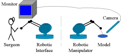
2.1. Telemanipulation System
 
The experiments use a laboratory teleoperation testbed consisting of two PHANTOM haptic interface devices (Model 1.5, SensAble Technologies, Inc., Woburn, Mass.). One PHANTOM acts as the surgeon master controller and the other acts as the surgical robot. The master is an unmodified PHANTOM with the stylus attachment. Subjects control the motion of the surgical robot by moving the stylus (Figure 1). 
The instrument used for the blunt dissection task is a right angle hook with a depth of 1 cm, a diameter of 0.9 mm, and a rounded tip. The hook is attached to a 50 cm rigid shaft that passes through a fixed pivot, simulating the incision into the patient's abdomen. The surgical robot is attached to the proximal end of the shaft with a two degree-of-freedom joint that prevents rotation of the instrument (Figure 2). 

 
Figure 1: Telemanipulation system

Figure 2: Surgical Setup

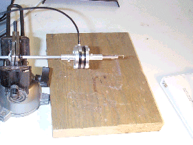
Figure 3: Hook and Force/Torque sensor
Forces are sensed at the tip of the instrument by a six-axis force/torque sensor (Mini transducer, ATI Industrial Automation, Apex, North Carolina) built into the instrument shaft (Figure 3). The PHANTOM control computer samples the forces at 1 kHz and transforms the forces to the proximal end of the shaft by assuming that the instrument shaft acts a perfect lever.  That force is scaled for the appropriate experimental condition and then reproduced by the surgeon master controller; ideally, this results in the user feeling the forces that would be experienced if the stylus was attached directly to the proximal end of the instrument shaft.
The teleoperation system, including the master, the surgical robot, and the force/torque sensor, are controlled by a 333 MHz Pentium computer running Windows NT.  The surgical robot's position is controlled using proportional position control, independent of force feedback.  The control algorithm is implemented in Visual C++ along with the force/torque sensor interface.

2.2. Visual Feedback
 
The subjects received visual feedback from a fixed  surgical endoscope, camera, and light source (Telecam SL NTSC/Xenon 175, Karl Storz Endoscopy-America, Inc., Culver City, Cal.), to provide the same visual feedback encountered in minimally invasive procedures. The relative orientation between the master controller and the monitor is approximately the same as orientation between the endoscope camera and the instrument, to minimize the mental effort of relating visual and instrument frames [15].  However, lack of depth perception and the laparoscopic movement constraint at the incision point remain sources of difficulty for untrained surgeons. 
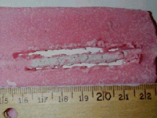
2.3. Surgical Models
 
The surgical models used here are intended to simulate a vital structure such as an artery embedded in its surrounding tissue. Two types of model were constructed: in one the artery was visible through the tissue and in the other tissue completely obscured the artery” These models contain materials of different stiffness on the order of the pertinent biological tissues to provide realistic stiffness contrast. Further, the models are straightforward to dissect with a fixed endoscopic view and an instrument with fixed orientation.
The material chosen to simulate the tissue bed is a clay similar to commercially available children's play dough.  The artery is represented by a stiffer clay material (Weatherstrip and Caulking Cord, Mortite, Inc., Kankakee, Ill.) in cylindrical strips 4 mm in diameter.  The tissue bed clay is colored pink to provide visual contrast to the gray artery material (Figure 4).  Although these clay materials do not simulate the viscoelastic characteristics of real tissue, they capture the plastic failure that is the goal of blunt dissection procedures. To quantify the material properties, we measured the steady dragging force of the blunt dissection hook embedded 5 mm into the model tissue material as 0.5 N, and embedded into the model artery material as 3.5 N.

 
Figure 4: Surgical Model

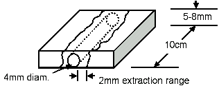
Figure 5: Model Schematic
A uniform and easily replicated fabrication process was used in the construction of these models. To fabricate each model, we placed a straight 10 cm length of artery on a mass of dough, then compressed the model with a flat plate to a uniform height.  For the model where the artery was visible, the tissue was compressed to a height of 5 mm.  For the obscured artery case, the model was compressed to 8 mm. The model was then flipped and squared off to regular dimensions, so that the artery was at the bottom of the resulting model (Figure 5). 

2.4. Experimental Setup
 
Subjects carried out several dissection tasks with varying levels of force feedback provided by the teleoperation system. Subjects were instructed to expose the artery, clearing away tissue 2 mm on each side of the artery as well as removing any tissue on top of the artery.  The subjects were also told to minimize the number of errors, defined as any scratch or puncture of the artery that exceeded a certain force threshold.  The threshold corresponding to visible damage of artery was found to be 0.25 N.  Aside from the primary goal of minimizing errors, the subject was instructed to minimize the area of tissue disturbed outside of the region to be exposed.  Finally, after meeting the above two requirements, the subject was to expose as much of the artery as possible in the allotted time. 
Figure 6: Exposed artery
In every case, the subject was to start at the same point and progress down the artery, working to clear both the sides and the top of the artery at the same time.  In the trials where the artery was not initially visible, the subjects were to find and then expose the artery (Figure 6). Subjects were informed that the artery was always generally straight and centrally located within the model.  Lastly, the subjects were to always use the same motion when clearing away tissue, that of a small scrape or dig with the hook instrument.
Subjects trained about 30 minutes in order to become familiar with the system and the task. Longer times were allowed for certain participants to ensure a similar level of proficiency across subjects. Each subject participated in 6 trials of 5 minutes each, where each trial involved a force feedback scaling of 0% (no force feedback), 75%, or 150%. Each of these force feedback levels was repeated for one model with a visible artery and one with an obscured artery. Eight subjects, 3 male and 5 female, participated in the study. All were students (mean age 25 years) with a basic familiarity with dissection.

2.5. Measures
 
Four different outcome measures were examined for each trial.  The applied forces, the number of errors, the length of dissection, and the area of tissue affected were chosen to best characterize the performance of a subject.  The applied forces, number of errors, and total area affected correlate directly with tissue trauma.  The length dissected, given a fixed time limit, provided a measure of productivity. 
All forces encountered by the instrument tip were recorded by the software.  To avoid recording force data when cleaning the tip of excess tissue, a button on the stylus was used to pause the logging of data. Peak and root-mean-square (RMS) force values were then extracted from the complete force record.   During the experiment, an observer noted each time the hook appeared to contact the artery as a possible error. The time of the possible error was recorded during the trial, and then verified by examining the force log at the time of the error and confirming that the force threshold was exceeded. Area affected was calculated using a digital image of the completed models.  The area affected was segmented from the image by hand and then measured using software.  Finally, length dissected was extracted using a similar method, using the digital image to measure the amount of artery exposed.

2.6 Statistical Analysis
 
For statistical analysis of the data the nonparametric Friedman test of k related samples was employed using the SPSS statistical analysis software package (Version 10.1, SPSS Inc., Chicago, Ill.).  The nonparametric test was chosen because of the relatively small sample and the lack of information concerning the distribution of the variables under study.  The statistical analysis included examining the RMS forces, the peak forces, the length dissected, the area affected per cm dissected, and the number of errors per trial.  A p value of less than 0.05 was considered statistically significant.


3. Results

Force feedback significantly reduced the magnitude of the forces applied at the instrument tip during dissection. Figure 7 shows a histogram of the force samples for all subjects and all trials with the visible artery; samples below 0.1 N are excluded. Subjects applied high force levels for longer durations when force feedback was not available. Conversely, during trials with force feedback, less time was spent applying higher forces; forces above 0.8 N were of negligible duration for 150% force feedback scaling, and above 1.2 N were negligible for 75% scaling. Further, the greater the force feedback gain, the less time was spent applying larger levels of force. These results also apply whether or not the subject can initially see the artery (Figure 8).


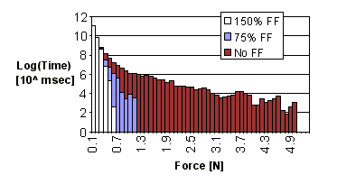
Figure 7: Histogram of forces applied during visible artery trials

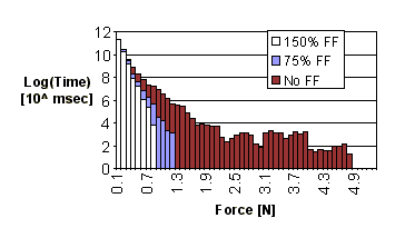
Figure 8:  Histogram of forces applied during obscured artery trials
Figures 9 and 10 show the RMS and peak forces under the two visibility conditions. The addition of force feedback significantly reduced the RMS force by 30% to 60% (visible artery, p = 0.008; obscured artery, p = 0.005) and the peak force by a factor of 2 to 6 (visible artery, p = 0.008; obscured artery, p = 0.002) Again, higher force feedback gain resulted in a reduction of forces applied. 
The average number of errors during a trial was also affected by the addition of force feedback (Figure 11).  Increased force feedback led to a significant (p = 0.001) reduction in the average errors, although increasing force scaling from 75 to 150% did not appreciably decrease the error rate.

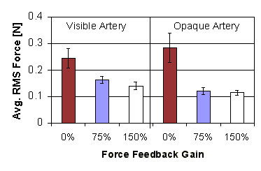
Figure 9: Average RMS force applied versus force feedback gain (error bars show standard error)

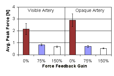
Figure 10: Average peak force applied versus force feedback gain (error bars show standard error)
Two measures that were not significantly affected by the addition of force feedback were the normalized length of artery dissected and the tissue area affected per cm of artery dissected (Figures 12, 13).  The lengths were normalized to the 0% force feedback/obscured artery case for each subject to reduce variations due to intersubject strategy differences. The length of artery dissected did not change significantly over different levels of force feedback or with the ability to see the artery in the tissue (artery visible, p = 0.093; artery obscured, p = 0.156).  Similarly, the amount of area dissected was not influenced by the addition of force feedback in either visual condition (p = 0.687 and 0.565 respectively).

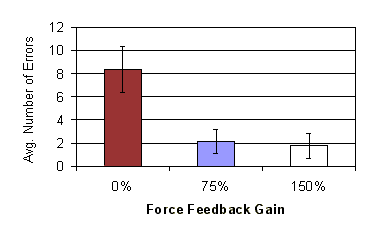
Figure 11: Average number of errors vs. force feedback gain (error bars show standard error)

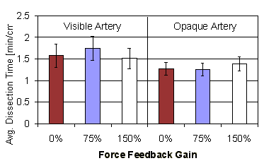
Figure 12: Normalized length dissected vs. force feedback gain (error bars show standard error)

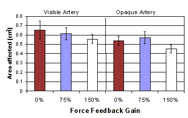
Figure 13: Area affected per cm dissected vs. force feedback gain (error bars show standard error)


4. Discussion
 
In this study we examine the effects of force feedback on a blunt dissection task, where we hypothesized that the addition of force feedback improves surgical performance. Our results show that force feedback improves performance by reducing the overall forces applied, thus reducing tissue trauma.  Force feedback also aids surgical performance by reducing the number of accidental incursions into sensitive structures. 
This study leads us to hypothesize that the benefits of force feedback to a surgeon are twofold (Figure 14).  At high levels of force feedback, we speculate that the intrinsic mechanical properties of the tissues being manipulated are transformed into physical constraints on the surgeon’s motions.  Subjectively speaking, it is difficult to move the instrument into a damaging configuration because a large force on the hand will oppose any motion that involves contact between the instrument and the tissue.  Further, this constraint not only acts as a safety barrier, reducing the forces applied and the number of errors, but the constraint can also act as a guide to the surgical instrument.  For instance, when the instrument is positioned between two structures of different stiffness, accurate dissection can simply be achieved by first applying a minimal force to press the instrument against the stiffer tissue. Then, the instrument can be dragged along the surface of the stiffer tissue while relying on the force feedback to maintain a uniform and safe contact force between tissue and instrument (Figure 15).
Figure 14: Hypothesized force feedback spectrum

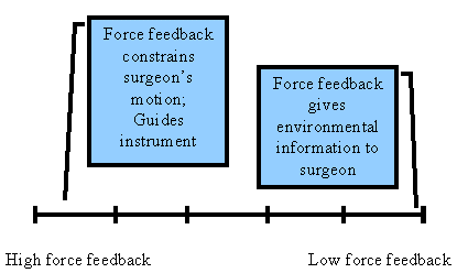
Figure 15: Instrument between two tissues of different stiffness
As the level of the provided force feedback decreases, the benefit of force feedback is hypothesized to arise less as a physical constraint and more as a supplemental source of information.  Because the forces are now harder to perceive, the surgeon must devote increased mental processing capacity to recognize and interpret this additional information.  Thus, at low levels of force feedback, a conscious response is required to take advantage of the available forces.  Further experimental investigation is required to understand the role of force magnitudes in this range.
Conditions in this study simulated the essential aspects of laparoscopic hook dissection in minimally invasive surgical procedures. Visual feedback was provided by a standard surgical laparoscope, and the instrument control mode included the fixed pivot at the incision point. While the mechanical properties of the synthetic clay models were not identical to actual tissue, the key behavior for this task is plastic deformation under traction loading. In this respect, the clay material faithfully replicates the behavior of tissue under blunt dissection with electrocautery. 
Based on informal experiments with real tissues, we conjecture that the relationships among force feedback gain and performance measures will persist over a range of mechanical properties; in particular, the same constraint mechanism functions in both cases. The variation in performance measured in this study as a function of force feedback gain were large and repeatable, and shown to a high degree of statistical significance. 
The subjects used in this study were novices, but planned future studies will use experienced surgeons for comparison. We assume that the lack of training in current subjects will have only a secondary effect on the outcome, because the results presented here suggest that the constraint generated by force feedback in this task appears to be readily used at all levels of training. Nonetheless, experienced laparoscopic surgeons have developed perceptual and motor skills to deal with the constraints of minimally invasive surgical techniques, and be able to use visual information to guide fine motions to avoid generating large forces. This visual approach would probably increase cognitive workload, however, so the aid provided by force feedback aid may decrease the likelihood of errors.
From this study, the benefit of force feedback is clear when accurate instrument positioning is required and/or when the involved structures are sensitive and trauma to the surrounding tissues has severe implications. Microsurgical procedures meet all these conditions and may be considered the likely candidates for dexterity enhancement by instruments with force feedback capabilities. Presently the visual acuity, dexterity and tactile sensitivity of the surgeon define the limits of microsurgical procedures.  The use of force feedback would allow scaling of forces up to perceivable levels, providing the aforementioned advantages to the microsurgical realm. 
Our future research efforts will first focus on verifying these results using subjects with surgical experience and biological tissues.  The next step will then be to further examine the role of force feedback in dissection with other types of dissectors.  One example would be the dissecting forceps, where pushing and spreading forces are encountered. There, we would expect to see similar benefits of safety and trauma reduction.  Additionally, we will examine the role of force feedback in the other main surgical tasks of cutting, grasping, retraction, and suturing.  In the case of grasping, adding force feedback to the grippers reduces tissue trauma and increases safety, and allows surgeons to tap into the human ability to sense slip to control grasp force [16]. Suturing is hypothesized to benefit from force feedback by allowing the mechanical tissue properties to guide and constrain the motion of the needle as it is being driven through a tissue plane. A final research goal is to quantify the effects of force feedback on mental workload in a surgical task.

5. References

[1] H. Shennib, A. Bastawisy, M. J. Mack, and F. H. Moll, "Computer-assisted telemanipulation: an enabling technology for endoscopic coronary artery bypass," Ann Thorac Surg, vol. 66, pp. 1060-3., 1998.
[2] E. R. Stephenson, Jr., S. Sankholkar, C. T. Ducko, and R. J. Damiano, Jr., "Robotically assisted microsurgery for endoscopic coronary artery bypass grafting," Ann Thorac Surg, vol. 66, pp. 1064-7., 1998.
[3] O. S. Bholat, R. S. Haluck, R. H. Kutz, P. J. Gorman, and T. M. Krummel, "Defining the role of haptic feedback in minimally invasive surgery," presented at Medicine Meets Virtual Reality. Convergence of Physical and Informational Technologies: Options for a New Era in Healthcare. MMVR 7, Amsterdam, Netherlands, 1999.
[4] A. Kazi, "Operator performance in surgical telemanipulation," Presence, vol. 10, pp. 495-510, 2001.
[5] J. Rosen, M. MacFarlane, C. Richards, B. Hannaford, and M. Sinanan, "Surgeon-tool force/torque signatures - evaluation of surgical skills in minimally invasive surgery," presented at Medicine Meets Virtual Reality. Convergence of Physical and Informational Technologies: Options for a New Era in Healthcare. MMVR 7, Amsterdam, Netherlands, 1999.
[6] A. K. Morimoto, R. D. Foral, J. L. Kuhlman, K. A. Zucker, M. J. Curet, T. Bocklage, T. I. MacFarlane, and L. Kory, "Force sensor for laparoscopic Babcock," Stud Health Technol Inform, vol. 39, pp. 354-61, 1997.
[7] P. J. Berkelman, L. L. Whitcomb, R. H. Taylor, and P. Jensen, "A miniature instrument tip force sensor for robot/human cooperative microsurgical manipulation with enhanced force feedback," presented at Medical Image Computing and Computer-Assisted Intervention - MICCAI 2000. Third International Conference. Proceedings, Berlin, Germany, 2000.
[8] A. Menciassi, A. Eisinberg, G. Scalari, C. Anticoli, M. C. Carrozza, and P. Dario, "Force feedback-based microinstrument for measuring tissue properties and pulse in microsurgery," presented at Proceedings 2001 ICRA. IEEE International Conference on Robotics and Automation, Piscataway, NJ, USA, 2001.
[9] S. Payandeh, "Force propagation models in laparoscopic tools and trainers," presented at Proceedings of the 19th Annual International Conference of the IEEE Engineering in Medicine and Biology Society. `Magnificent Milestones and Emerging Opportunities in Medical Engineering', Piscataway, NJ, USA, 1997.
[10] J. Rosen, B. Hannaford, M. P. MacFarlane, and M. N. Sinanan, "Force controlled and teleoperated endoscopic grasper for minimally invasive surgery-experimental performance evaluation," IEEE Transactions on Biomedical Engineering, vol. 46, pp. 1212-21, 1999.
[11] M. C. Cavusoglu, A. Sherman, and F. Tendick, "Bilateral controller design for telemanipulation in soft environments," presented at Proceedings 2001 ICRA. IEEE International Conference on Robotics and Automation, Piscataway, NJ, USA, 2001.
[12] T. B. Sheridan, Telerobotics, automation, and human supervisory control. Cambridge, Mass.: MIT Press, 1992.
[13] R. S. Johansson and G. Westling, "Signals in tactile afferents from the fingers eliciting adaptive motor responses during precision grip," Exp Brain Res, vol. 66, pp. 141-54, 1987.
[14] C. E. H. Scott-Conner, The SAGES manual: fundamentals of laparoscopy and GI endoscopy. New York: Springer, 1999.
[15] F. Tendick, R. Jennings, G. Tharp, and L. Stark, "Sensing and Manipulation Problems in Endoscopic Surgery," Presence, vol. 2, pp. 66-81, 1993.
[16] B. B. Edin, R. D. Howe, G. Westling, and M. Cutkosky, "A physiological method for relaying frictional information to a human teleoperator," IEEE Transactions on Systems, Man and Cybernetics, vol. 23, pp. 427-432, 1993.
 

Back to Past Projects